
2019 - 2 Issue

Review

Normal Tension vs High Tension Glaucoma: An Overview

Full Text links

Summary

The study provides an up-to-date overview of pathogenesis, functional and structural changes in normal tension glaucoma (NTG) and its differences from high tension glaucomas (HTG).

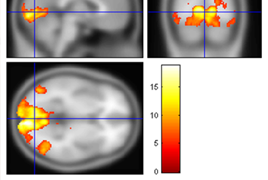
The authors point to less known facts which make both diagnostic groups different. First of all, there are electrophysiological findings that verify pathology in the complete visual pathway in HTG in contrast to NTG where the retinal ganglion cell response is relatively normal but the abnormalities are in the visual pathway. This corresponds to the findings of functional magnetic resonance imaging of the brain with a significant decrease in activity in HTG compared to NTG. We found a higher decrease in activity in HTG following application of the colour paradigm compared to NTG where we did not see a similar difference. We also investigated the central corneal thickness (CCT) in both diagnostic groups. We did not find a statistically significant difference. However, we found the effect of CCT on progression of the changes in visual fields in HTG. In relation to suspicion of abnormally low cerebrospinal pressure and a possible cerebrovascular fluid flow disturbance in NTG, we examined the optic nerve thickness (OND) and optic nerve sheath diameter (OSD) at a distance of 4, 8, 16 and 20mm from the posterior pole of the eye. In the comparison with the healthy population, we did not find any abnormalities except for the width of the optic chiasma that was markedly lower in NTG. In relation to a possible impairment of cerebral perfusion we determined the degrees of cerebral atrophy using magnetic resonance imaging by measuring the bicaudate ratio (BCR) and white matter lesions using the Fazekas scale. We did not find a difference between HTG and NTG in BCR. We found statistically significant changes in BCR which correlated with the changes in visual fields. The higher values of the pattern defect were associated with increased brain atrophy (BCR). We did not detect similar relations in the Fazekas scale. We found a significant difference in this parameter among NTG, HTG and a control group. We found the most advanced changes in the patients with HTG.

Conclusion: In HTG, impairment of retinal ganglion cells and subsequently also their axons, including visual cortex occurs because of a high intraocular pressure. In NTG, the retinal ganglion cells are relatively normal like the visual cortex, but alteration occurs in their axons. The cause is not a high intraocular pressure but most probably ischemia.

References

  1. Araie, M., Yamagami, J., Suziki, Y.: Visual field defects in normal-tension and high-tension glaucoma. Ophthalmology, 100; 1993: 1808–1814.
  2. Bonomi, L., Marchini, G., Marraffa, M. et al.: Prevalence of glaucoma and intraocular pressure distribution in a defined population. The Egna-Neumarkt Study. Ophthalmology, 105; 1998: 209-215.
  3. Cecchi, E., Liotta, A., Gori, AM. et al.: Comparison of hemorheological variables in ST-elevation myocardial infarction versus those in non-ST-elevation myocardial infarction or unstable angina pectoris. Am J Cardiol, 102; 2008: 125-128.
  4. Flammer, J., Konieczka, K.: The discovery of the Flammer syndrome: a historical and personal perspective. EPMA Journal, 8; 2017: 75-97.
  5. Flammer, J., Prünte, C.: Ocular vasospasm. 1: Functional cir - culatory disorders in the visual system, a working hypothesis. Klin Monbl Augenheilkd, 198; 1991: 411–412.
  6. Fortune, B., Bui, BV., Morrison, JC. et al.: Selective ganglion cell functional loss in rats with experimental glaucoma. Invest Ophthalmol Vis Sci, 45; 2004: 1854-1862.
  7. Hamard, P., Hamard, H., Dufaux, J. et al.: Optic nerve head blood flow using a laser Doppler velocimeter and haemorheology in primary open angle glaucoma and normal pressure glaucoma. Br J Ophthalmol, 78; 1994: 449-453.
  8. Hayreh, SS.: Pathogenesis of cupping of the optic disc. Br J Ophthalmol, 58; 1974: 863-876.
  9. Holder, GE.: Pattern electroretinography (PERG) and an integrated approach to visual pathway diagnosis. Prog Retin Eye Res, 20; 2001: 531-561.
  10. Chang, M., Yoo, C., Kim, SW. et al.: Retinal Vessel Diameter, Retinal Nerve Fiber Layer Thickness, and Intraocular Pressure in Korean Patients with Normal-Tension Glaucoma. Am J Ophthalmol, 151; 2011: 100-105.
  11. Cheng, HC., Chan, CM., Yeh, SI. et al.: The Hemorheological Mechanisms in Normal Tension Glaucoma. Curr Eye Res, 36; 2011: 647-653.
  12. Cho, HK., Kee, C.: Population-based glaucoma prevalence studies in Asians. Surv Ophthalmol, 59; 2014: 434-447.
  13. Khodabandehlou, T., Le Dévéhat, C.: Hemorheological disturbances as a marker of diabetic foot syndrome deterioration. Clin Hemorheol Microcirc, 30; 2004: 219-223.
  14. Killer, HE., Miller, NR., Flammer J, et al.: Cerebrospinal fluid exchange in the optic nerve in no.rmal-tension glaucoma. Br J Ophthalmol, 96; 2012: 544-548.
  15. Klein, BE., Klein, R., Sponsel, WE. et al.: Prevalence of glaucoma. The Beaver Dam Eye Study. Ophthalmology, 99; 1992: 1499-1504.
  16. Lester, M., De Feo, F., Douglas, GR.: Visual field loss mor - phology in high-and normal-tension glaucoma. J Ophthalmol, 2012; 27326. Epub 2012: Feb 8.
  17. Lestak, J., Jiraskova, N., Zakova, M. et al.: Normotensive glaucoma. Biomed Pap Med Fac Univ Palacky Olomouc Czech Repub, 162; 2018: 272-275.
  18. Lešták, J., Nutterová, E., Pitrová, Š. et al.: High tension versus normal tension glaucoma. A comparison of structural and functional examinations. J Clinic Exp Ophthalmol, 2012; S5:006. doi:10.4172/2155-9570.S5-006
  19. Lešták, J., Pitrová, Š., Nutterová, E.: Centrální tloušťka rohovky u normotenzních a hypertenzních glaukomů. Cesk Slov Oftalmol, 74; 2018: 186-189.
  20. Lešták, J., Rozsíval, P.: The Influence of Corneal Thickness on Progression of Hypertensive Glaucoma. J Clin Exp Ophthal - mol, 2012; 3: 245. doi:10.4172/2155-9570.1000245
  21. Lestak, J., Tintera, J., Svata, Z. et al.: Glaucoma and CNS. Comparison of fMRI results in high tension and normal tension glaucoma. Biomed Pap Med Fac Univ Palacky Olomouc Czech Repub, 158; 2014: 144–153.
  22. Lin, PW., Friedman, M., Lin, HC. et al.: Normal tension glaucoma in patients with obstructive sleep apnea/hypopnea syndrome. J Glaucoma, 20; 2011: 553-558.
  23. Lindén, C., Qvarlander, S., Jóhannesson, G. et al.: Normal- Tension Glaucoma Has Normal Intracranial Pressure: A Prospective Study of Intracranial Pressure and Intraocular Pressure in Different Body Positions. Ophthalmology, 125; 2018: 361-368.
  24. Nebbioso, M., Gregorio, FD., Prencipe, L. et al.: Psychophysiological and electrophysiological testing in ocular hypertension. Optom Vis Sci, 55; 2011: 928-939.
  25. Okuno, T., Sugiyama, T., Kojima, S. et al.: Diurnal variation inmicrocirculation of ocular fundus and visual field change in normal-tension glaucoma. Eye (Lon), 18; 2004: 697–702.
  26. Parisi, V., Miglior S., Manni, G. et al.: Clinical ability of pattern electroretinograms and visual evoked potentials in detecting visual dysfunction in ocular hypertension and glaucoma. Ophthalmology, 113; 2006: 216-228.
  27. Plange, N., Remky, A., Arend, O.: Colour Doppler imaging and fluorecein fillingdefects of the optic disc in normal tension glaucoma. Br J Ophthalmol, 87; 2003: 731–736.
  28. Rotchford, AP., Johnson, GJ.: Glaucoma in Zulus: a population-based cross-sectional survey in a rural district in South Africa. Arch Ophthalmol, 120; 2002: 471-478.
  29. Shin, IH., Kang, SY., Hong, S. et al.: Comparison of OCT and HRT findings among normal, normal tension glaucoma, and high tension glaucoma. Korean J Ophthalmol, 22; 2008: 236–241.
  30. Shin, JW., Sung, KR., Lee, JY. et al.: Optical coherence tomography vessel density mapping at various retinal layers in healthy and normal tension glaucoma eyes. Graefes Arch Clin Exp Ophthalmol, 255; 2017: 1193-1202.
  31. Shou, T., Liu, J., Wang, W. et al.: Differential dendritic shrinkage of alpha and beta retinal ganglion cells in cats with chronic glaucoma. Invest Ophthalmol Vis Sci, 44; 2003: 3005- 3010.
  32. Schwenn, O., Troost, R., Vogel, A. et al.: Ocular pulse amplitude in patients with open angle glaucoma, normal tension glaucoma, and ocular hypertension. Br J Ophthalmol, 86; 2002: 981–984.
  33. Velcheva, I., Antonova, N., Titianova, E. et al.: Hemorheological disturbances in cerebrovascular diseases. Clin Hemorheol Microcirc, 39; 2008: 391-396.