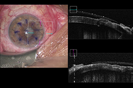
Optical coherence tomography (OCT) is a non-contact and non-invasive imaging and diagnostic method, that allows the imaging of ocular tissues on transverse sections in extremely high quality of micrometer resolution. The physical principle of OCT is analogous to ultrasound, but it uses infrared radiation instead of acoustic waves. By using a low coherent radiation source, it is possible to achieve a higher resolution. Based on the obtained data, the computer can reconstruct two or three-dimensional images of the examined tissue. In recent years, we have seen a rapid development in ophthalmic surgery, especially in surgical instruments and imaging methods. However, the technology of surgical microscopes does not change significantly and thus becomes a limiting factor in the development of ophthalmic microsurgery. The integration of the OCT into surgical microscopes, so the introduction of the Intraoperative Optical Coherence Tomography (iOCT), opens up further possibilities for displaying/looking at the operating field and adapting surgical techniques to the current situation during the surgery. On the contrary, the disadvantage is the prolongation of the surgery time and thus the theoretical increase in possible complications related to the surgery. iOCT can be used for operations on the anterior and posterior segment of the eye. In anterior segment surgery, it is used mostly in penetrating and lamellar keratoplasty, glaucoma surgery and cataract surgery. In posterior segment surgery, it is used during pars plana vitrectomy. The aim of the article is to provide a comprehensive overview of the current possibilities of using iOCT in eye surgery.
- Intraoperative Optical Coherence Tomography –Available Technologies and Possibilities of Use. A Review
- Laser Vitreolysis in Patients with Symptomatic Vitreous Floaters
- Tube Versus Trabeculectomy in Juvenile-Onset Open Angle Glaucoma – Treatment Outcomes in Tertiary Hospitals In Malaysia
- Assessment of Corneal Endothelial Layer in Contact Lens Wearers with the Aid of an Endothelial Microscope
- Tubulointerstitial Nephritis With Uveitis (Tinu Syndrome). A Case Report
- Late Choroidal Neovascular Complications in a Patient Treated for Retinoblastoma. A Case Report