Aim: To report a case of progressive corneal thinning and spontaneous corneal perforation in the setting of recurrent keratoconjunctivitis of undetermined etiology in a 20-year-old female patient.
Material and methods: Longitudinal follow-up of the reported case was performed with regular slit-lamp examination, anterior segment optical coherence tomography, and laboratory evaluation. Relevant scientific literature was reviewed to discover potential etiologies and causes of the reported case.
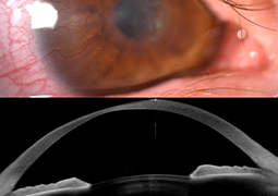
Case report: Recurrent bilateral keratoconjunctivitis was detected and followed up in a 20-year-old female patient. Long-standing blepharitis and several ocular inflammatory episodes were observed involving the conjunctiva and cornea causing corneal thinning and irregular astigmatism on both eyes. Patient’s history, physical and laboratory examination did not reveal any systemic inflammatory or dermatological disorder. After 4 years from the onset of symptoms, spontaneous corneal perforation was observed on the right eye and treated with amniotic membrane transplantation. At that time, eyelid margin culture was positive for Streptococcus mitis. The graft healed completely with paracentral stromal scarring. Best spectacle corrected visual acuity was 20/25 OD and 20/40 OS a month after the procedure.
Conclusion: Corneal and ocular surface inflammation is a potential multifactorial disease. Bacterial hypersensitivity, atopy and dermatological disorders such as atopic dermatitis and rosacea may play a role in recurrent keratitis, corneal thinning and eventually corneal perforation. Frequent and regular follow-ups are required to detect complications early as well as to discover all possible local and systemic contributing factors of keratoconjunctivitis.