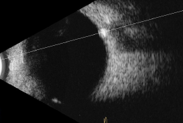
Aim: The aim of this retrospective study is to compare two methods of optic disc drusen imaging in pediatric patients – new swept source OCT technology with the B-scan ultrasonography, which has been assumed to be a gold standard in diagnosing optic disc drusen, and to compare pRNFL thickness in children with optic disc drusen and control group.
Methods: 14 eyes of 8 pediatric patients in whom optic disc drusen were confirmed by either B-scan ultrasonography, or swept-source OCT, were included in the study. We compared the sensitivity of these two imaging methods. Then we compared an average pRNFL thickness and pRNFL thickness in all four quadrants of our study group with the control group. Two statistical methods were used in data analysis – Mann-Whitney a Kruskal-Wallis test.
Results: The difference between SS-OCT and B-scan ultrasonography was not statistically significant in diagnosing optic disc drusen.
Average pRNFL thickness was 135.29 ± SD 31.2 μm in eyes with optic disc drusen, which is 24.15 % higher than in control group (p = 0.00214; p = 0.00207).
pRNFL thickness of temporal (p = 0.0001; p = 0.0001), superior (p = 0.03486; p = 0.03361) and inferior (p = 0.00652; p = 0.00627) quadrant was statistically significantly higher in comparison with healthy controls, whereas the difference of pRNFL thickness in nasal quadrant was not statistically significant between the study and control group (p = 0.09692; p = 0.0947).
Conclusion: Swept source OCT is a promising new "gold standard" in optic disc drusen diagnostics in pediatric patients. An increase in pRNFL thickness values does not always confirm papilloedema as optic disc drusen may be the cause. Direct visualisation of optic disc drusen by swept source OCT can aid in differentiation from true papilloedema.