Aim: To clarify the possibilities and role of posterior segment imaging in patients with neurofibromatosis type I (NF1), and to show the prevalence of this disease in the pediatric population in Slovakia.
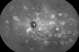
Material and methods: Until recently, ophthalmologic consultations in patients with NF1 were limited mainly to the observation of Lisch nodules of the iris and the presence of optic nerve glioma. However, advances in imaging capabilities have made it possible to investigate and describe new findings concerning the ocular manifestations of this disease. Between October 2020 and November 2021, we examined the anterior and posterior segment of 76 eyes (38 children – 12 boys and 26 girls) with genetically confirmed NF1 gene mutation at our clinic. The age of the patients ranged from 4 to 18 years. The anterior segment was checked for the presence of Lisch nodules biomicroscopically with a slit lamp. On the posterior segment, the presence of choroidal nodules was checked by various imaging methods – fundus camera, infrared confocal selective laser ophthalmoscopy, MultiColor imaging, OCT, and OCT angiography. All the patients had magnetic resonance imaging performed in order to detect potential optic nerve gliomas for the purpose of diagnosis. We observed the correlation between the patients’ age, presence of Lisch nodules and the presence of choroidal nodules. Eight patients also had other manifestations of the disease – optic nerve gliomas or microvascular changes (so-called “corkscrew” vessels).
Results: Out of 38 patients, Lisch iris nodules were present in 20 patients (53%) and choroidal nodules in 24 patients (63%). There was no positive correlation between the presence of these two manifestations within the same patient or eye, but there is a clear correlation between the presence of choroidal nodules and patient age.
Conclusion: The results suggest that a previously unknown ocular manifestation of neurofibromatosis type I, namely choroidal nodules, has a higher prevalence than Lisch nodules also in the pediatric population and can be easily visualized using various imaging modalities. It will be important to include follow-up observation of this finding among the standard controls for ocular findings in NF1, and it will be very interesting to correlate this finding with the exact NF1 mutation.