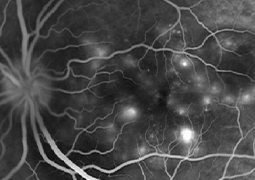
Aims: Vogt-Koyanagi-Harada (VKH) disease is an autoimmune disorder affecting multiple systems and characterized by bilateral granulomatous uveitis, frequently together with neurological, auditory, and integumentary manifestations. Prompt diagnosis and intervention are a must to prevent irreversible vision loss and possible systemic complications. Fundus imaging plays a pivotal role in the management of VKH, not only to reach the diagnosis but also to monitor disease activity and response to treatment. Fundus photography, fundus autofluorescence imaging, fluorescein angiography, indocyanine green angiography, and optical coherence tomography (OCT) are among the imaging modalities. These methods provide invaluable insights into the different phases of the disease. In addition, OCT angiography enables clinicians to visualize associated retinal and choroidal microvascular changes. Continuous advances in imaging technology assist ophthalmologists to comprehend the VKH pathophysiology and to facilitate its differential diagnosis. Thus, clinical outcomes are improving with timely interventions and personalized treatment approaches. This mini-review provides an overview of VKH disease, with a particular focus on the fundus imaging techniques utilized in its management.
- Use of Imaging Modalities in Vogt-Koyanagi-Harada Disease: An Overview
- Rhegmatogenous Retinal Detachment in Childhood
- Reconstruction of the Anophthalmic Conjunctival Sac. A Review of Surgical Procedures to Achieve Stability of the Ocular Prosthesis in Our Practice
- Focusing on the Future: Patient-Centered Insights into Trifocal Intraocular Lens Adoption
- Presentation of a New Method for Quantitative Determination of Trifocal Intraocular Lens Decentration
- Recurrent Congenital Cytomegalovirus Chorioretinitis in a Newborn. A Case Report