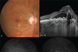
Progress in optical coherent tomography (OCT) has recently provided new insights into variety of chorioretinal disorders. The use of EDI (Enhanced Depth Imaging) during OCT examinations and as well as OCT angiography provides a more accurate analysis of the choroid both quantitatively and qualitatively. „Pachychoroid“ (greek pachy- [παχύ]: thick) is defined as an abnormal and sustained increase in choroidal thickness ≥ 300 μm, which is mainly due to dilated choroidal vessels in the Haller’s layer and other structural changes in physiological choroidal architecture. Central serous chorioretinopathy is one of many macular diseases associated with „pachychoroid“. Another diseases that belong to the group of macular pachychoroid disorders are: pachychoroid pigment epitelopathy, pachychoroid neovasculopathy, polypoid choroidal vasculopathy. In this paper we summarize the current view on pachychoroid macular diseases and describe characteristics that are observed in multimodal imaging analysis of choroidal changes.
- Pachychoroid disease of the macula
- Use of EX-PRESS® implant in glaucoma surgery - retrospective study
- Change of tear osmolarity after refractive surgery
- Orbital optic nerve sheath meningioma
- Femtosecond laser - Assisted intrastromal corneal segment implantation – our experience
- Merkel cell carcinoma of the eyelid and orbit