
2024 - 6 Issue

Original article

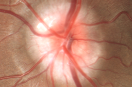
Evaluation of Optic Disc Drusen Using Modern Imaging Paraclinical Methods

Full Text links

Summary

Purpose: To analyze patients with optic disc drusen (ODD), with emphasis on modern diagnostics.

Materials and Methods: Research of the literature was conducted, together with a retrospective statistical analysis of patients with ODD. The group included individuals with ODD diagnosed using at least one of the following (ultrasound – USG, optical coherence tomography – OCT, fundus autofluorescence – FAF).

Results: The group consisted of 12 patients (23 eyes), 7 women and 5 men. The mean age was 25 years. The mean observation period was 73 months. In total, 11 patients (22 eyes) had a bilateral finding and 1 patient (1 eye) had a unilateral finding. The mean age was 25 years. Buried drusen were confirmed in 69.6% of cases (8 patients, 16 eyes), superficial drusen were confirmed in 30.4% of cases (4 patients, 7 eyes). Mean best corrected visual acuity (BCVA) and mean intraocular pressure were stable over time (BCVA p = 0.236, IOP p = 0.855). The aforementioned diagnostic methods proved to be equally effective (p = 0.768). In 11 patients (21 eyes) a depression of the retinal nerve fiber layer (RNFL) was recorded. We found a statistically significant decrease of the RNFL over time in reference to the normative database in the superior temporal (p = 0.015), temporal (p = 0.026) and nasal segments (p = 0.011). After separation of superficial and buried drusen the same significant change was found in nasal segment in superficial drusen (p = 0,031). We found no statistically significant difference over time between superficial and buried drusen (p = 0.109–0.999 for individual segments).

Conclusion: ODD are common and visual functions remain stable. Their presence can be confirmed using modern paraclinical methods.

References

  1. Friedman AH, Beckerman B, Gold DH, Walsh JB, Gartner S. Drusen of the optic disc. Surv Ophthalmol. 1977;21(5):375-390.
  2. Tso MO. Pathology and pathogenesis of drusen of the optic nervehead. Ophthalmology. 1981;88(10):1066-1080.
  3. Palmer E, Gale J, Crowston JG, Wells AP. Optic Nerve Head Drusen: An Update. Neuro-ophthalmology. 2018;42(6):367-384.
  4. Thurtell MJ, Biousse V, Bruce BB, Newman NJ. Optic nerve head drusen in black patients. J Neuroophthalmol. 2012;32(1): 13-16.
  5. Rosenberg MA, Savino PJ, Glaser JS. A clinical analysis of pseudopapilledema. I. Population, laterality, acuity, refractive error, ophthalmoscopic characteristics, and coincident disease. Arch Ophthalmol. 1979;97(1):65-70.
  6. You QS, Xu L, Wang YX, Jonas JB. Prevalence of optic disc drusen in an adult Chinese population: the Beijing Eye Study. Acta Ophthalmol. 2009;87(2):227-228.
  7. Mansour AM, Hamed LM. Racial variation of optic nerve diseases. Neuro-ophthalmology. 1991;11(6):319-323.
  8. Štrofová H, Jarošová A. Drúzy papily zrakového nervu a jejich komplikace [Optic Disc Drusen and their Complications]. Cesk Slov Oftalmol. 2016;72(1):298-308. Czech.
  9. Obuchowska I, Mariak Z. Zaburzenia pola widzenia w druzach tarczy nerwu wzrokowego [Visual field defects in the optic disc drusen]. Klin Oczna. 2008;110(10-12):357-360. Polish.
  10. Nentwich MM, Maertz J, Rudolph G. Drusenpappile – ein Überblick über medizinhistorische und aktuelle Aspekte [Optic Disk Drusen: Historical and Up-To-Date Aspects]. Klin Monbl Augenheilkd. 2016;232(3):257-265. German.
  11. Jirásková N, Rozsíval P. Dekomprese obalů zrakového nervu [Decompression of the optic nerve sheath]. Cesk Oftalmol. 1995;51(4):254-257. Czech.
  12. Jirásková N, Rozsíval P. Dekomprese obalů zrakového nervu – výsledky prvních 37 operovaných očí [Decompression of the optic nerve sheath-results in the first 37 operated eyes]. Cesk Slov Oftalmol. 1996;52(5):297-307. Czech.
  13. Jirásková N, Rozsíval P. Výsledky 62 dekompresí obalů zrakového nervu [Results of 62 optic nerve sheath decompressions]. Cesk Slov Oftalmol. 1999;55(3):136-144. Czech.
  14. Grippo TM, Shihadeh WA, Schargus M, et al. Optic nerve head drusen and visual field loss in normotensive and hypertensive eyes. J Glaucoma. 2008;17(2):100-104.
  15. Nolan KW, Lee MS, Jalalizadeh RA, Firl KC, Van Stavern GP, McClelland CM. Optic Nerve Head Drusen: The Relationship Between Intraocular Pressure and Optic Nerve Structure and Function. J Neuroophthalmol. 2018;38(2):147-150.
  16. Tuğcu B, Özdemir H. Optik disk druseni Tanisinda Görüntüleme Yöntemleri [Imaging Methods in the Diagnosis of Optic Disc Drusen]. Turk J Ophthalmol. 2016;46(5):232-236. Turkish.
  17. Hamann S, Malmqvist L, Costello F. Optic disc drusen: understanding an old problem from a new perspective. Acta Ophthalmol. 2018;96(7):673-684.
  18. Kurz-Levin MM, Landau K. A comparison of imaging techniques for diagnosing drusen of the optic nerve head. Arch Ophthalmol. 1999;117(8):1045-1049.
  19. Denniston AKO, Murray PI. Oxford Handbook of Ophthalmology. 3rd ed. Oxford (England): Oxford University Press; 2014. Chapter 2, Investigations and their interpretation; p. 63.
  20. Pfriem M, Hoerauf H. Unsuccessful surgical excision of optic nerve drusen. Graefes Arch Clin Exp Ophthalmol. 2011;249(10):1583- 1585.
  21. Kapur R, Pulido JS, Abraham JL, Sharma M, Buerk B, Edward DP. Histologic findings after surgical excision of optic nerve head drusen. Retina. 2008;28(1):143-146.
  22. Roh S, Noecker RJ, Schuman JS. Evaluation of coexisting optic nerve head drusen and glaucoma with optical coherence tomography. Ophthalmology. 1997;104(7):1138-1144.
  23. Gramer G, Gramer E, Weisschuh N. Optic Disc Drusen and Family History of Glaucoma-Results of a Patient-directed Survey. J Glaucoma. 2017;26(10):940-946.
  24. Auw-Haedrich C, Staubach F, Witschel H. Optic disk drusen. Surv Ophthalmol. 2002;47(6):515-532.
  25. Suelves AM, Francés-Muñoz E, Gallego-Pinazo R, et al. Central serous papillopathy by optic nerve head drusen. Clin Ophthalmol. 2010;4:1379-1382.+